anatomy inner ear ct
The Radiology Assistant : Anatomy 2.0. 9 Pictures about The Radiology Assistant : Anatomy 2.0 : Middle ear anatomy: Stapes, facial nerve canal, oval and round window, 3D CT Middle and Inner Ear and also Superior semicircular canal dehiscence | Image | Radiopaedia.org.
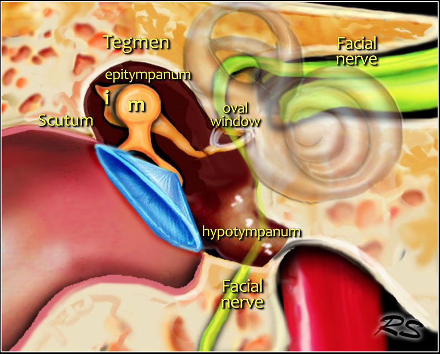
The Radiology Assistant : Anatomy 2.0
 radiologyassistant.nl
radiologyassistant radiology assistant tympanic cavity
The Radiology Assistant : Temporal Bone - Anatomy 2.0 In 2021
 www.pinterest.com
radiology
Otic Capsule: Annotated CT | Radiology Case | Radiopaedia.org
 radiopaedia.org
capsule otic ct radiopaedia annotated radiology labyrinth case ear cochlea
Middle Ear Anatomy: Stapes, Facial Nerve Canal, Oval And Round Window
 www.researchgate.net
temporal ear oval anatomy stapes
Superior Semicircular Canal Dehiscence | Image | Radiopaedia.org
 radiopaedia.org
semicircular canal superior dehiscence ct radiopaedia version
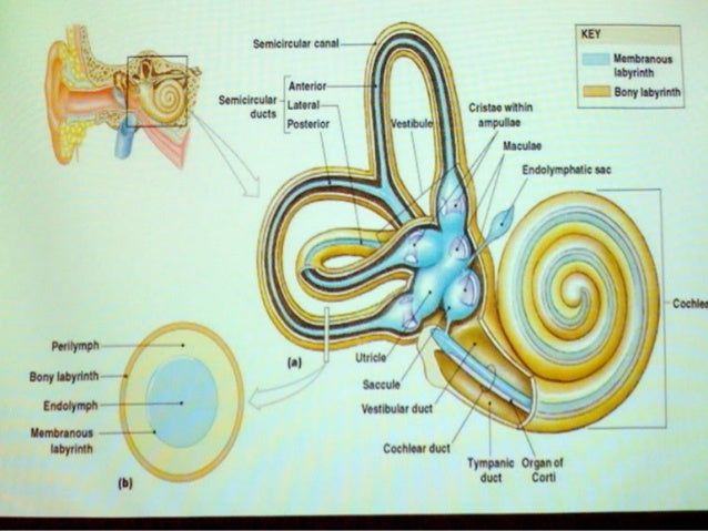
INNER EAR - DR NITIN ANIYAN THOMAS (NATS)
 www.slideshare.net
ear anatomy vestibule endolymph perilymph bony duct membranous corti crus canals tympanic cochlear aniyan nitin
3D CT Middle And Inner Ear
 www.slideshare.net
ear ct middle inner 3d slideshare scan
Facial Nerve Anatomy - Labeled CT | Radiology Case | Radiopaedia.org
 radiopaedia.org
facial ct nerve labeled anatomy radiopaedia
Cholesteatoma - Wikidoc
 www.wikidoc.org
cholesteatoma wikidoc symptoms ear
The radiology assistant : temporal bone. The radiology assistant : anatomy 2.0. 3d ct middle and inner ear