anatomy female abdominal
Midcarpal joint: Anatomy | Kenhub. 9 Images about Midcarpal joint: Anatomy | Kenhub : Yoga for Spine Mobility: Anatomy of the Spine and Rib Cage | Yoga, Normal female pelvis, MRI - Stock Image C026/9010 - Science Photo Library and also Abdominal Wall and Intestines CADAVER (Dr. Rawa) - YouTube.
Midcarpal Joint: Anatomy | Kenhub
wrist pollicis joint adductor bones carpal midcarpal anatomy kenhub muscle retinaculum grip flexor musculus hand anterior insertion origin capsule ventral
Schwannoma Of The Nasal Cavity: Clinical Case, Diagnosis | Kenhub
nasal cavity case sagittal clinical nasopharynx section kenhub conchae hard middle schwannoma anatomy soft mid cadaveric inferior alqu
Normal Female Pelvis, MRI - Stock Image C026/9010 - Science Photo Library
 www.sciencephoto.com
mri pelvis normal female sciencephoto
Superior Gluteal Artery: Anatomy, Banches, Supply | Kenhub
superior gluteal artery kenhub anatomy extremity arterial glutea arteria lower anastomoses supply dorsal
Abdominal Wall And Intestines CADAVER (Dr. Rawa) - YouTube
 www.youtube.com
cadaver intestines abdominal lab dr
K.Mayotte Rad Tech | Rad 2325 Group 1 Disscussion Forum
 openlab.citytech.cuny.edu
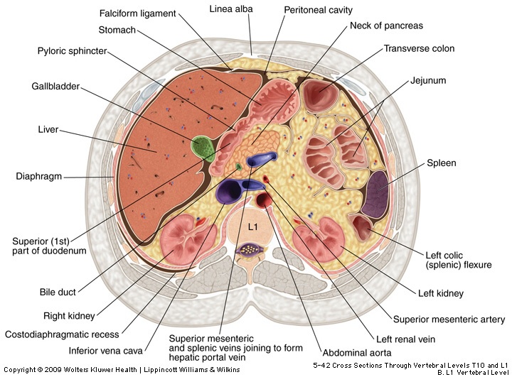
section cross anatomy cholelithiasis stomach kidneys lower superior ct gallbladder kidney spleen scan limb blood edu duct rad mayotte tech
Musculocutaneous Nerve: Anatomy, Course And Function | Kenhub
nerve musculocutaneous kenhub course anatomy nervus function ventral branches
Yoga For Spine Mobility: Anatomy Of The Spine And Rib Cage | Yoga
 www.pinterest.com
rib cage anatomy muscles muscle spine ribs human body yoga envy thoracic mobility bones visit yogajournal source
Epiglottis: Structure, Function, Epiglottitis | Kenhub
epiglottis kenhub anatomy function structure epiglottitis pharyngeal dorsal
Yoga for spine mobility: anatomy of the spine and rib cage. Epiglottis kenhub anatomy function structure epiglottitis pharyngeal dorsal. Nasal cavity case sagittal clinical nasopharynx section kenhub conchae hard middle schwannoma anatomy soft mid cadaveric inferior alqu