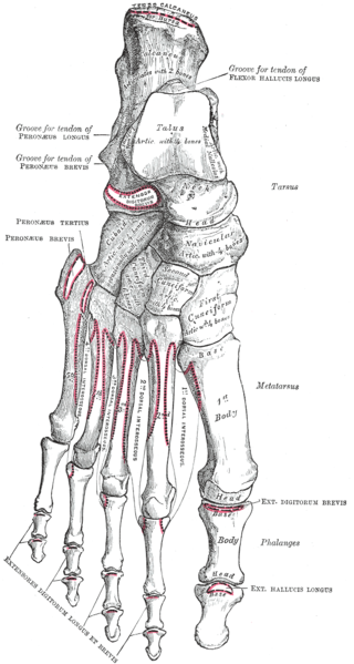
anatomy bones of foot
Shoulder Radiographic Anatomy in 2021 | Radiology student, Radiology. 9 Pictures about Shoulder Radiographic Anatomy in 2021 | Radiology student, Radiology : Foot, Plantar Surface (deeper) – Human Body Help, Surface Anatomy - Atlas of Anatomy and also Arches of the foot: Anatomy | Kenhub.
Shoulder Radiographic Anatomy In 2021 | Radiology Student, Radiology
 www.pinterest.com
anatomy shoulder lateral radiographic scapular radiology position medical oblique student joint wikiradiography ray positioning schools axillary normal radiograph neers anterior
Foot, Plantar Surface (deeper) – Human Body Help
 www.humanbodyhelp.com
foot plantar surface deeper key
Ankle Bones, Talus, Navicular, Cuneiforms, Calcaneus, And Cuboid
 www.pinterest.com
talus appendicular navicular cuboid calcaneus cheville cuneiforms tarsus tarsal naviculaire tarse cuboïde visiblebody squelette anatomie consists
Chopart Fracture-dislocation - Physiopedia
 www.physio-pedia.com
chopart foot bones fracture dislocation epidemiology etiology
Arches Of The Foot: Anatomy | Kenhub
foot arch arches longitudinal kenhub lateral anatomy right tissues soft
Anatomy Of The Foot Medical Illustration Medivisuals
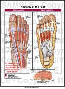 medivisuals1.com
foot anatomy 01x plantar dorsal right medical medivisuals1 illustration
Knee Posterior View
 www.thinglink.com
knee posterior
Surface Anatomy - Atlas Of Anatomy
 doctorlib.info
anatomy surface medical where atlas hepatic portal branching easynotecards doctorlib info superior triple down
Historical Anatomies On The Web: William Cheselden Home
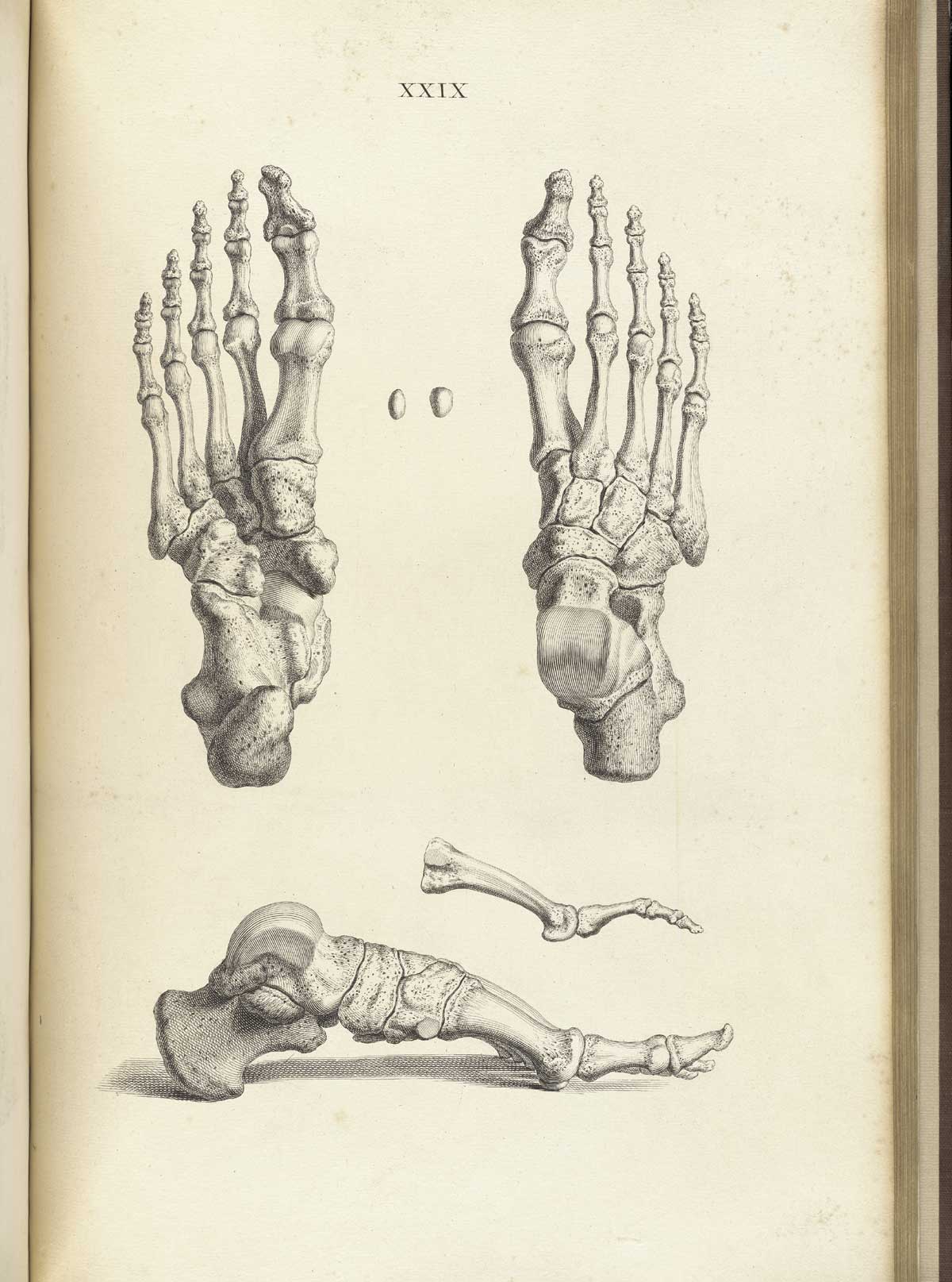 www.nlm.nih.gov
anatomy bones skeleton foot drawing cheselden human william nlm feet osteographia skull nih gov historical bone bottom left side drawings
Knee posterior view. Anatomy of the foot medical illustration medivisuals. Chopart fracture-dislocation