anatomy bone femur
Pelvic X-Ray Anatomy and Interpretation Checklist | GrepMed. 9 Images about Pelvic X-Ray Anatomy and Interpretation Checklist | GrepMed : The Hip Joint - Complete Physiotherapy, Third trochanter ( "Occasionally" present on femur ) - Gives attachment and also Hüftgelenk - Anatomie, Aufbau, Knochen, Bänder & Muskeln | Kenhub.
Pelvic X-Ray Anatomy And Interpretation Checklist | GrepMed
 www.grepmed.com
pelvis pelvic xray grepmed
Intraosseous Lipoma Of The Proximal Femur | Image | Radiopaedia.org
 radiopaedia.org
lipoma intraosseous femur proximal radiopaedia version radiology case bone
The Hip Joint - Complete Physiotherapy
 www.completephysiotherapy.co.uk
hip joint anatomy human bones body replacement ball resurfacing bone pelvis femur skeletal where system virtual diagram ligament spine conditions
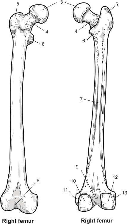
Print Pelvis & Femur Flashcards | Easy Notecards
 www.easynotecards.com
femur
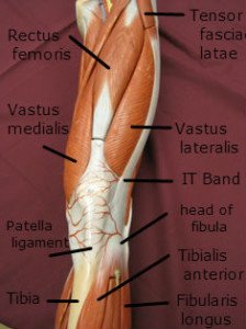
Iliotibial Band (IT Band) Attachment
 chiropractor-sioux-city.com
iliotibial attachment anatomy quadriceps peroneus longus fibularis enthesopathy graston tendinitis ligaments ligament pelvis muscular chandler sioux chiropractor
Hüftgelenk - Anatomie, Aufbau, Knochen, Bänder & Muskeln | Kenhub
Eosinophilic Granuloma Of The Femur | Image | Radiopaedia.org
 radiopaedia.org
eosinophilic femur granuloma ray langerhans histiocytosis cell bone radiology chest pathology radiopaedia wikidoc orthobullets 1036 case version
Third Trochanter ( "Occasionally" Present On Femur ) - Gives Attachment
 in.pinterest.com
femur trochanter tendon tendonitis bursitis tendons ligaments trochanteric gluteus beaconortho bony piriformis rib sheena johns
Femur Fracture Before And After Surgery Stock Photo & More Pictures Of
 www.istockphoto.com
fracture femur surgery broken before bones anatomy
Iliotibial band (it band) attachment. Iliotibial attachment anatomy quadriceps peroneus longus fibularis enthesopathy graston tendinitis ligaments ligament pelvis muscular chandler sioux chiropractor. Femur fracture before and after surgery stock photo & more pictures of