3d knee anatomy
Systematic Interpretation of Knee MRI: How I do it - ViYoutube. 9 Images about Systematic Interpretation of Knee MRI: How I do it - ViYoutube : {Knee Joint showing image of the meniscus along with the Cruciat, Foot - Superficial and deep dissection of distal leg and foot | 3D and also Transient Lateral Patellar Dislocation - Radsource.
Systematic Interpretation Of Knee MRI: How I Do It - ViYoutube
 viyoutube.co
mri systematic tendon
Foot - Superficial And Deep Dissection Of Distal Leg And Foot | 3D
 www.ahealthcare.com
distal foot leg dissection superficial deep
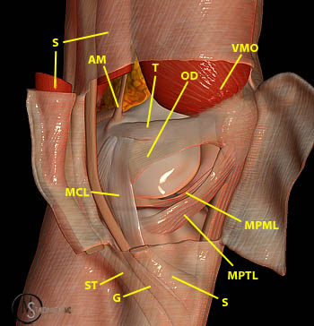
{Knee Joint Showing Image Of The Meniscus Along With The Cruciat
 johnthebodyman.com
knee joint meniscus ligaments posterior inferior along showing anatomy previous
Acute Pain In Knee Stock Images - Image: 30511194
 dreamstime.com
pain acute knee isolated man background
Anatomy Of The Canine Knee - EasyAnatomy
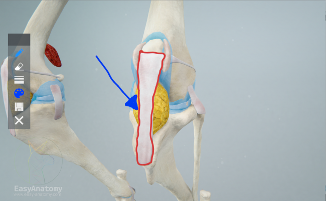 easy-anatomy.com
canine patellar anatomy ligament knee easy
Transient Lateral Patellar Dislocation - Radsource
 radsource.us
patellar lateral dislocation mri radsource transient
Full Body Pain Anatomy — Stock Photo © Pixologic #22672903
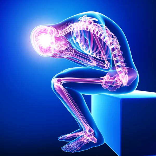 depositphotos.com
pain body male anatomy joint highlighted skeleton depositphotos illustration organs inner system pixologic circulatory
Posterolateral Corner Injury - Radsource
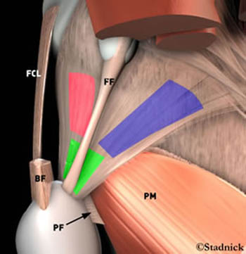 radsource.us
collateral ligament knee mri fibular corner posterolateral arcuate lateral injury popliteus fcl muscle radsource ligaments 2003 orthobullets structures skeletal musculo
Facial Artery: Anatomy, Branches And Clinical Points | Kenhub
artery facial anatomy lateral facialis branches left kenhub
Posterolateral corner injury. Artery facial anatomy lateral facialis branches left kenhub. Distal foot leg dissection superficial deep